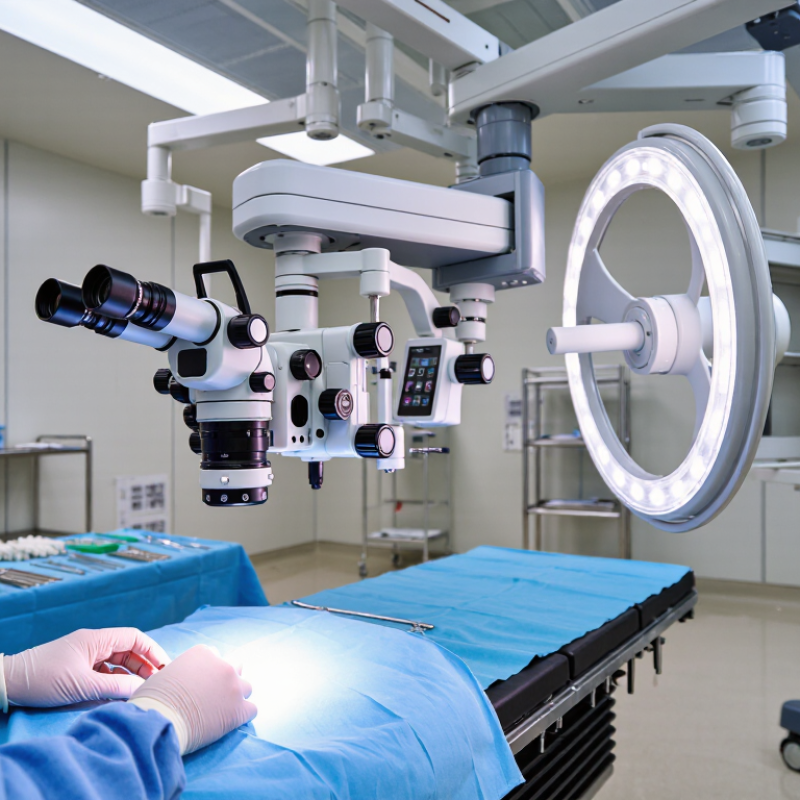
| Conventional optical surgical microscope | Direct binocular viewing through eyepieces; optical magnification and illumination support detailed work in a small operative field. | The surgeon generally looks through oculars. Assistant viewing depends on the configuration and may require an assistant scope or other accessories. | Familiar workflow and direct optical viewing can suit microsurgical tasks such as nerve-root and dural work. | Check working distance, field of view, illumination, focus controls, movement range, and whether the viewing posture is comfortable for the intended approach. | Teams that prefer eyepiece-based viewing and need a microscope for established microsurgical workflows. |
| Motorized optical microscope | Uses optical binocular viewing; motorized movement or focus functions may be available, depending on the configuration. | Motorized positioning can reduce some manual adjustments. Ergonomic benefit depends on controls, balance, reach, and how the microscope is set up. | Can support frequent repositioning across a surgical field, including during procedures that require changing viewing angles. | Verify which movements are motorized, how controls are operated, the time needed to reposition, and compatibility with the operating-room layout. | Surgeons who value optical viewing but want powered positioning or focus features. |
| Digital 3D exoscope | Displays a magnified surgical view on a monitor; 3D depth perception is available in systems designed for stereoscopic viewing and requires compatible viewing equipment. | The surgeon and team can view the same monitor image. A screen-based workflow may allow a more upright head and neck posture, depending on monitor placement. | Shared visualization can make it easier for the surgical team to follow the field; may suit teaching and procedures where screen viewing is preferred. | Assess image latency, monitor position, depth perception, focus and zoom controls, lighting, and performance at the working distance required for spine procedures. | Teams seeking shared screen-based visualization and a potential alternative to prolonged eyepiece viewing. |
| Hybrid optical and digital system | Combines optical viewing with a digital display or recording pathway, where included in the configuration. | May offer eyepiece viewing for the primary surgeon and a monitor view for other team members; available options vary. | Can accommodate mixed preferences for direct viewing and shared display, without requiring every team member to use the same viewing method. | Clarify whether the digital image is simultaneous, what accessories are required, and whether switching between viewing modes interrupts workflow. | Departments with surgeons or procedures that benefit from both eyepiece and monitor-based viewing. |
| Image-guided microscope integration | May combine microscope visualization with navigation or imaging information displayed in the field or on a separate screen; integration depends on compatible equipment. | Can bring relevant visual information into the team’s workflow, but may add setup steps and require coordination among systems. | Potentially useful when the procedure already uses navigation or other image-guidance tools; it does not replace clinical judgment or standard verification steps. | Confirm supported interfaces, registration workflow, display location, setup time, staff training needs, and what happens if integration is unavailable. | Spine teams that routinely use compatible navigation or imaging systems and can support the added workflow. |