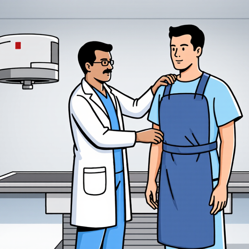
| What X-rays are | X-rays are high-energy electromagnetic radiation and are classified as ionizing radiation. | They can remove electrons from atoms and may damage cellular DNA. | Medical imaging should use the lowest radiation exposure that provides adequate diagnostic information. |
| Average natural background exposure | Approximately 3 mSv per year worldwide, although the amount varies by location and altitude. | This is ongoing low-level exposure from cosmic rays, soil, rocks, and naturally occurring radioactive materials. | A lead apron is not normally used for natural background radiation; exposure reduction focuses on limiting unnecessary medical exposure. |
| Dental X-ray | A typical intraoral dental X-ray is about 0.005 mSv, but the dose varies by equipment and technique. | The small exposure contributes mainly to a very low potential lifetime cancer risk. | Collimation, digital detectors, correct positioning, and avoiding repeat images are important. Apron policies may vary by local regulations and professional guidance. |
| Chest X-ray | A typical two-view chest X-ray is approximately 0.1 mSv, with variation by patient size and technique. | The radiation dose is low, but repeated or unnecessary examinations should still be avoided. | Proper beam collimation and exposure settings are primary controls. A lead apron may protect areas outside the useful beam when appropriate. |
| Mammography | A typical two-view screening examination is approximately 0.4 mSv, depending on the system and breast characteristics. | The main concern from low-dose exposure is a small increase in lifetime stochastic risk. | Breast compression and optimized imaging settings help reduce dose while maintaining image quality. |
| Head CT scan | A typical adult head CT is approximately 2 mSv, although protocols and patient size can change the dose. | The principal concern at this level is a small potential increase in long-term cancer risk; tissue reactions are not expected from a routine scan. | A lead apron cannot shield the brain from the CT beam. Protocol optimization and avoiding unnecessary repeat scans are more effective. |
| Abdomen and pelvis CT | A typical adult abdomen-and-pelvis CT is about 10 mSv, with substantial variation by protocol and patient size. | Several organs receive radiation, so avoiding unjustified examinations and repeat scans is important. | A lead apron placed over the scanned region cannot meaningfully reduce dose from the primary CT beam and may interfere with automatic exposure control. |
| Scatter radiation | Scatter is produced when the primary X-ray beam interacts with the patient or other materials and changes direction. | Repeated occupational exposure to scattered radiation can accumulate over time. | Lead aprons are mainly useful for reducing scatter exposure to workers or patients who must remain near the X-ray source. |
| Lead-equivalent apron thickness | Protective aprons are commonly specified by lead equivalence, such as 0.25 mm or 0.5 mm Pb, depending on the intended use. | Greater lead equivalence generally provides greater attenuation, but protection depends on X-ray energy, angle, coverage, and proper fit. | An apron must fully cover the area at risk, be inspected for cracks, and be stored without sharp folds or creases. |
| Time, distance, and shielding | Radiation protection follows three basic principles: minimize time, maximize distance, and use suitable shielding. | Reducing exposure time and increasing distance can substantially lower occupational dose. | The apron is one layer of protection and should be used together with barriers, distance, controlled exposure time, and dosimetry when required. |
| Possible biological effects | Effects are broadly divided into stochastic effects and tissue reactions. | Stochastic effects, such as cancer, are probability-based. Tissue reactions, such as skin injury, generally require much higher localized doses. | Routine diagnostic X-ray examinations are not expected to cause skin burns, but prolonged fluoroscopy or interventional procedures require strict protection and monitoring. |
| Pregnancy considerations | The fetus is more sensitive during certain stages of development, particularly at higher doses. | The potential effect depends on fetal dose, gestational age, and the area examined. | Patients should inform healthcare professionals about known or possible pregnancy. Necessary diagnostic imaging should not automatically be delayed. |